Anatomical Drawings Brain
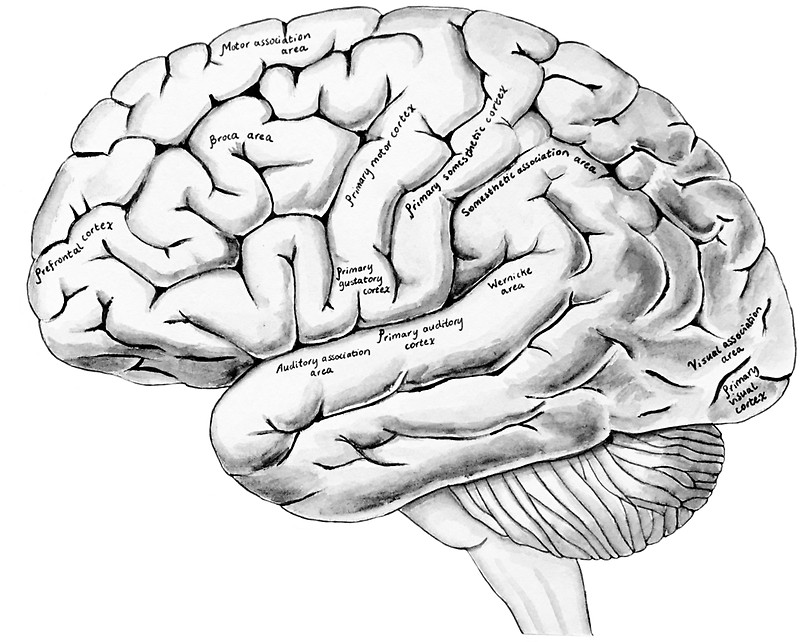
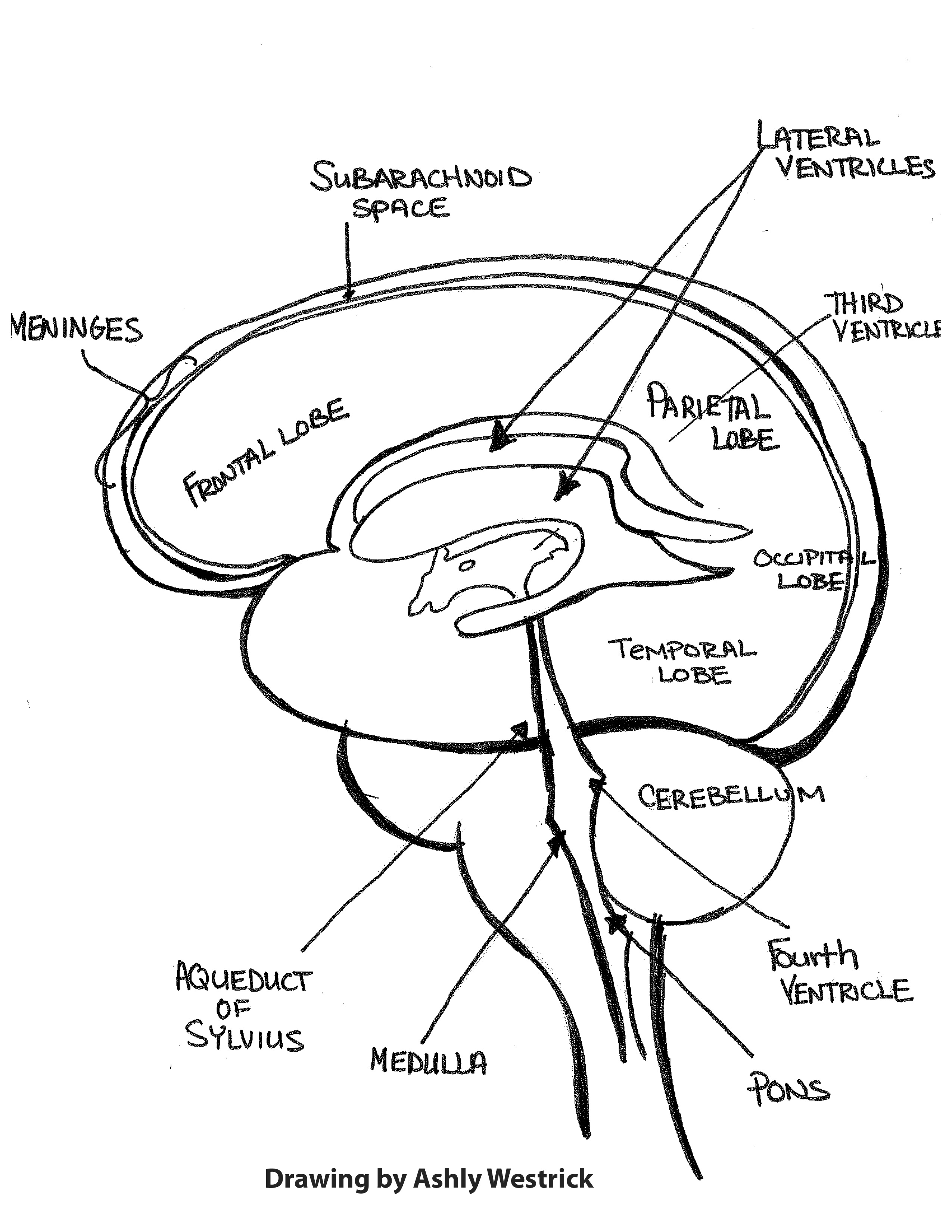
Anatomical Drawings Brain - The cerebrum, diencephalon, cerebellum, and brainstem. Web engage your kids in the world of brain anatomy with simple brain drawings. This interactive brain model is powered by the wellcome trust and developed by matt wimsatt and jack simpson; Web to gain a good knowledge of the brain through brain drawing is a great way to better understand anatomy and its connection to the brain. Web working from three images from the fabrica —a skeleton, a figure of muscles, and an illustration of the brain—this exhibit shows the many ways vesalius’ work built on past anatomists, and exerted its influence well into the future. Web between 1487 and 1493 leonardo created a number of marvelous drawings of the skull. The illustrations are labeled in three categories: Their illustrations, illustrators, and methods are discussed. Web by methodically integrating the cerebellum into your brain illustration, you enhance the comprehensive portrayal of the brain's anatomy, showcasing the intricate structures that underpin essential motor functions and coordination. This fun and educational activity will help them develop their artistic skills and provide them with valuable knowledge about the brain's anatomical structures. The brain has three main parts: Web the brain illustrations of vesalius and willis were the first in anatomic history with pictorial accuracy. Web by reflecting the innovative methods he applied to his research, leonardo's anatomical drawings can be considered among the most inspiring documents providing evidence of the empirical origins of modern scientific method. Understanding the brain through a brain line drawing or a diagrammatic method can also break down the different components of the brain and can help us better understand its complex composition. If you’re struggling, you might benefit from taking a few quizzes on the anatomy of the brain (see below). The brain directs our body’s internal functions. Explore the brain's complex functions and composition with innerbody's 3d anatomical model. Web there are 25 woodcut figures of brains, reflected dura, skulls, and vessels in andreas vesalius' landmark anatomic text the fabric of the human body (latin: Reviewed by john morrison, patrick hof, and edward. Web working from three images from the fabrica —a skeleton, a figure of muscles, and an illustration of the brain—this exhibit shows the many ways vesalius’ work built on past anatomists, and exerted its influence well into the future. Web the brain is a complex organ that controls thought, memory, emotion, touch, motor skills, vision, breathing, temperature, hunger and every process that regulates our body. The brain is one of the most complex and magnificent organs in the human body. Web engage your kids in the world of brain anatomy with simple brain drawings. These will allow you to. Reviewed by john morrison, patrick hof, and edward. Woodcut blocks were used for the prints of figures in the vesalian anatomy. Web “cells of the brain” presents some of the basics, beginning with pyramidal neurons, and including the pericellular nests that surround them like pointy hats, or eva hesse sculpture, and. The brain has three main parts: Web the brain. The cerebrum, diencephalon, cerebellum, and brainstem. It also integrates sensory impulses and information to form. Web we created a brain atlas that is an interactive tool for studying the conventional anatomy of the normal brain based on a magnetic resonance imaging exam of the axial brain. Figures of the brain appear to be done after external fixation in the work. If you want more of a challenge, include anatomical parts, such as the brain stem and cerebellum. Web between 1487 and 1493 leonardo created a number of marvelous drawings of the skull. Web the brain illustrations of vesalius and willis were the first in anatomic history with pictorial accuracy. Web these anatomical charts include the main diagrams necessary for medical. If you’re struggling, you might benefit from taking a few quizzes on the anatomy of the brain (see below). The user views the anatomical drawings by using the illustrations menu where each item corresponds to a cranial nerve. If you want more of a challenge, include anatomical parts, such as the brain stem and cerebellum. Web using this atlas of. Rotate this 3d model to see the four major regions of the brain: Woodcut blocks were used for the prints of figures in the vesalian anatomy. Web it's not always easy remembering the parts of the brain. If you want more of a challenge, include anatomical parts, such as the brain stem and cerebellum. Web between 1487 and 1493 leonardo. Web engage your kids in the world of brain anatomy with simple brain drawings. Web the brain illustrations of vesalius and willis were the first in anatomic history with pictorial accuracy. Anatomical structures and specific areas are. Web “cells of the brain” presents some of the basics, beginning with pyramidal neurons, and including the pericellular nests that surround them like. You can make it as simple as you like by drawing lots of squiggles and keeping the shape round. Web here, we’ve gathered some of our favorite historical anatomical drawings, which medieval and early modern doctors made from dissections of both animals and human cadavers. Our brain gives us awareness of ourselves and of our environment, processing a constant stream. The brain is one of the most complex and magnificent organs in the human body. Web “cells of the brain” presents some of the basics, beginning with pyramidal neurons, and including the pericellular nests that surround them like pointy hats, or eva hesse sculpture, and. If you want more of a challenge, include anatomical parts, such as the brain stem. Together, the brain and spinal cord that extends from it make up the central nervous system, or cns. Web we created a brain atlas that is an interactive tool for studying the conventional anatomy of the normal brain based on a magnetic resonance imaging exam of the axial brain. Web using this atlas of human anatomy. Rotate this 3d model. Figures of the brain appear to be done after external fixation in the work of willis. Web engage your kids in the world of brain anatomy with simple brain drawings. The user views the anatomical drawings by using the illustrations menu where each item corresponds to a cranial nerve. 1 the identity of the artist was who did the illustrations is uncertain. These will allow you to identify and work on your weak spots. Reviewed by john morrison, patrick hof, and edward. Web we created a brain atlas that is an interactive tool for studying the conventional anatomy of the normal brain based on a magnetic resonance imaging exam of the axial brain. If you want more of a challenge, include anatomical parts, such as the brain stem and cerebellum. Web the brain is one of the most fun parts of the body to draw. Cranial nerves, foramina and organs. Our brain gives us awareness of ourselves and of our environment, processing a constant stream of sensory data. Woodcut blocks were used for the prints of figures in the vesalian anatomy. The cerebrum is the largest and most recognizable part of the brain. This interactive brain model is powered by the wellcome trust and developed by matt wimsatt and jack simpson; Anatomical structures and specific areas are. Web by methodically integrating the cerebellum into your brain illustration, you enhance the comprehensive portrayal of the brain's anatomy, showcasing the intricate structures that underpin essential motor functions and coordination.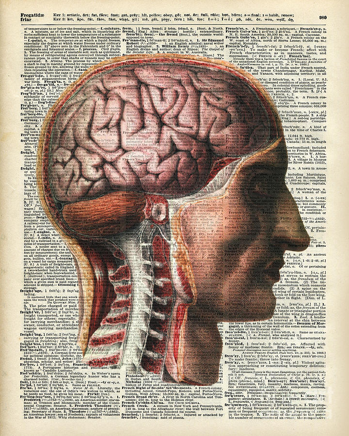
Vintage Human Brain Anatomy Drawing by Anna Wilkon

Brain drawing, Anatomy art, Brain art
Anatomical Brain hand drawn organ sketch. Medicine, Vector illustration
Reticulum Labeled Anatomical Vector Illustration Diagram Learning
brainanatomy Rachel Gold
Normal Anatomy of Human Brain Vintage Print 2 Drawing by Vintage
Anatomical Brain Drawing at GetDrawings Free download
Anatomical Brain Drawing at GetDrawings Free download

Anatomical Brain Drawing

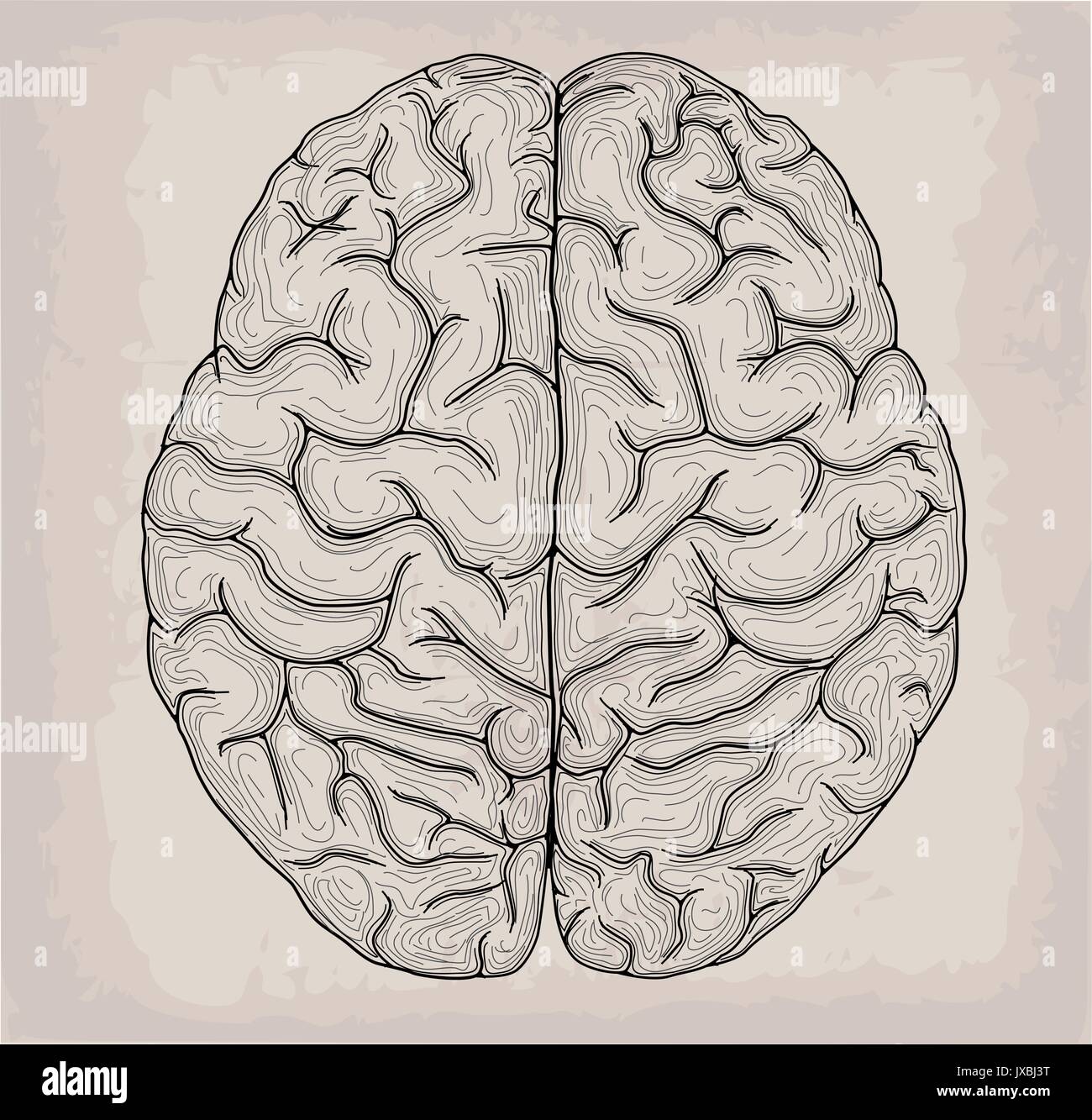
Human anatomy drawings brain front and top Vector Image
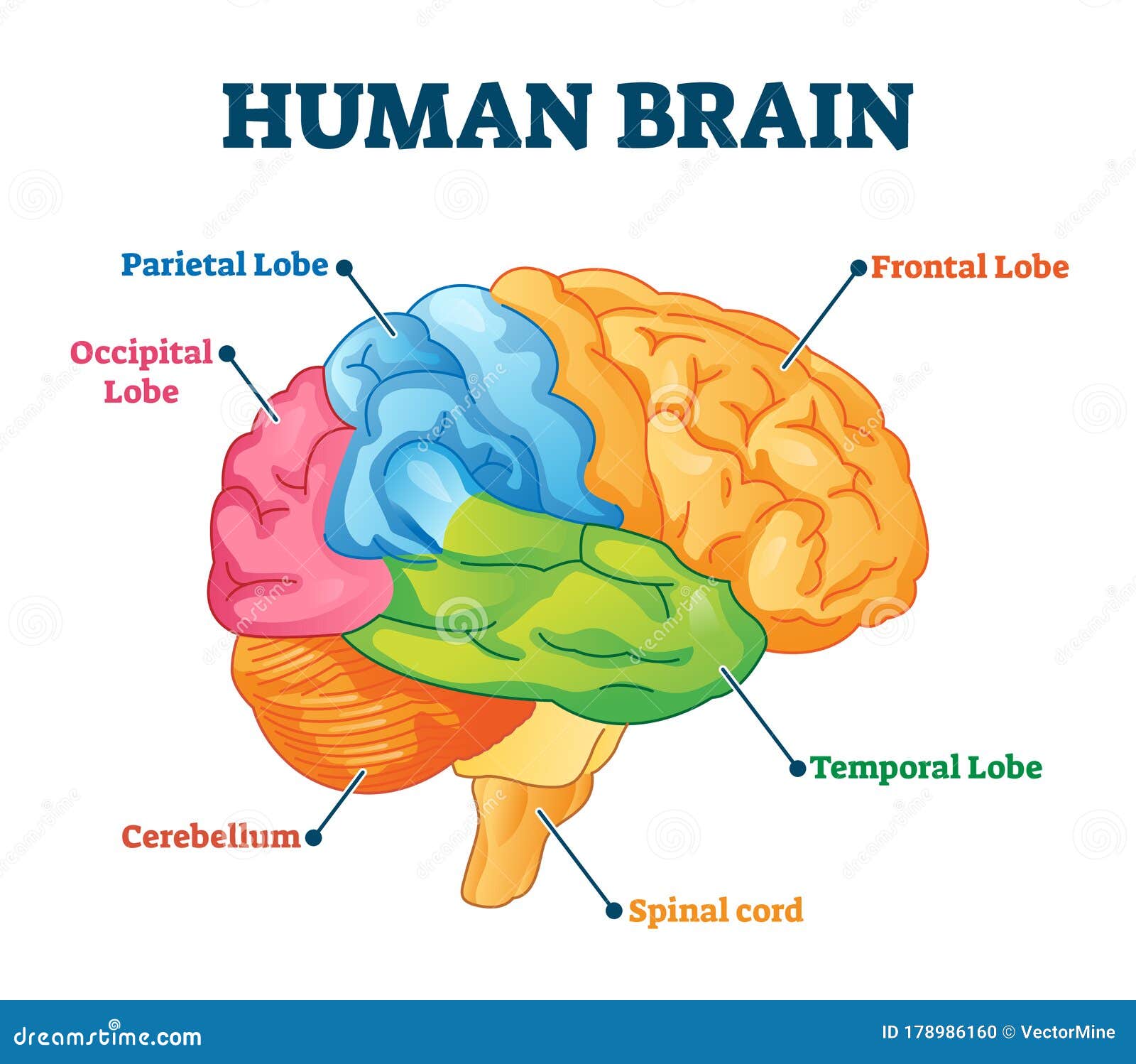
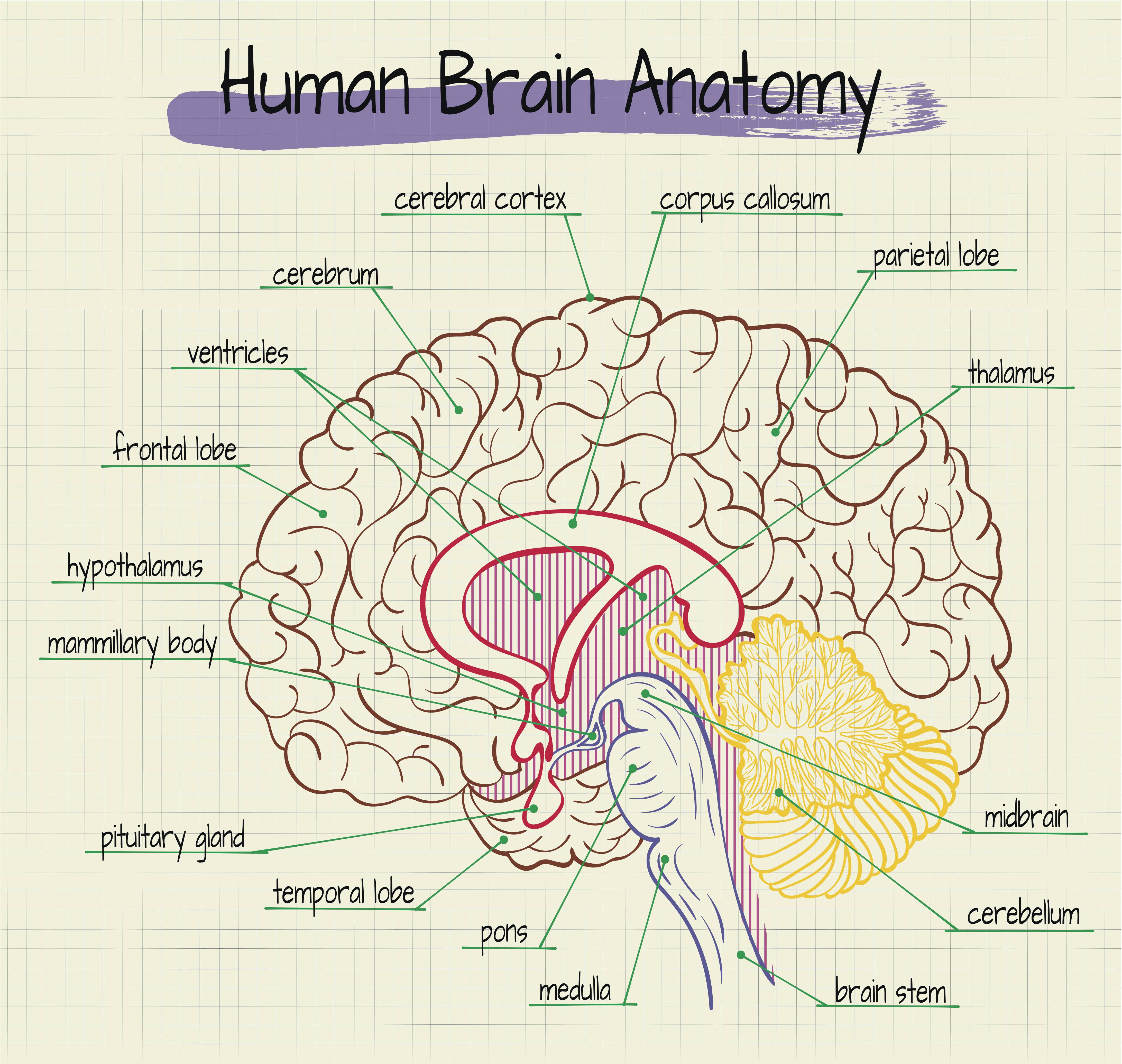
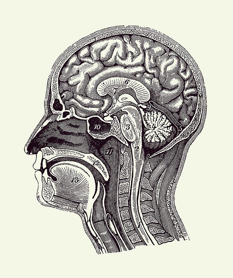
The Cerebrum, Diencephalon, Cerebellum, And Brainstem.
Web Between 1487 And 1493 Leonardo Created A Number Of Marvelous Drawings Of The Skull.
Web To Gain A Good Knowledge Of The Brain Through Brain Drawing Is A Great Way To Better Understand Anatomy And Its Connection To The Brain.
Web These Anatomical Charts Include The Main Diagrams Necessary For Medical Students, Nursing Students, Residents, Practitioners, Anatomists To Study The Anatomy Of The Brain, To Illustrate A Course Or Explain A Pathology To A Patient.
Related Post: